
Each year, MS Australia holds two grant rounds to select only the top MS research projects to fund. Further information about the comprehensive grant review process is available here.
The 19 new projects announced in 2026 address MS Australia’s priorities for MS research, including causes and prevention, better treatments and cures via repair and regeneration of cells. The projects are driving groundbreaking research in key areas, from using cutting-edge technologies to identifying biomarkers for earlier MS detection, developing innovative treatments for motor symptoms, promoting myelin repair, and exploring lifestyle impacts.
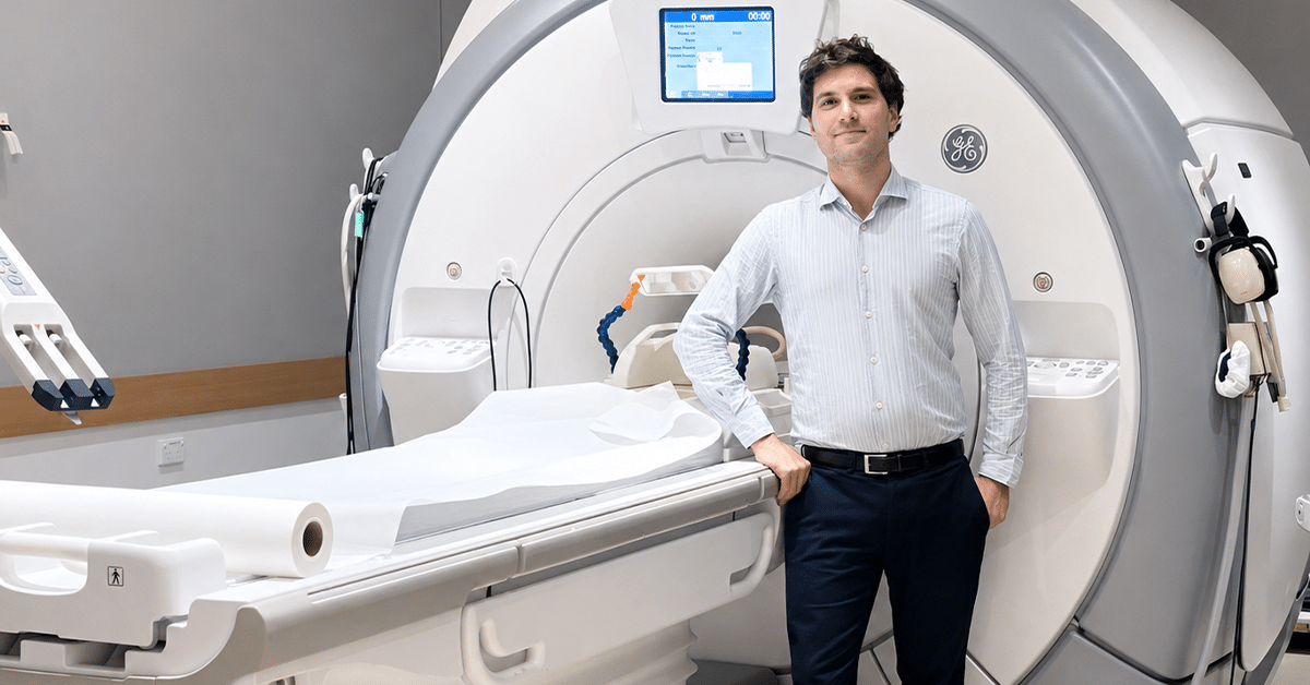
Multiple sclerosis (MS) can damage the spinal cord. This damage is strongly linked to physical disability and disease progression. However, current magnetic resonance imaging (MRI) scans are not sensitive enough to reliably detect small changes in the spinal cord over time. This makes it harder for doctors to accurately monitor disease activity and progression. Dr Koren says people with MS often express frustration that their MS is worsening but there is no clear evidence of this on their MRI scan. This project aims to improve how spinal cord damage is detected and measured in people with MS.
Dr Tal Koren will analyse existing MRI scans from people with MS who have had spinal cord imaging over several years. He will measure areas of damage and changes in spinal cord size over time. These scans will be used to develop and train an artificial intelligence (AI) model that can automatically detect and track spinal cord damage across multiple timepoints.
He will then recruit adults with MS who are already undergoing routine MRI scans. In addition to the standard scans, participants will receive a newer, high-resolution 3D MRI scan of the spinal cord. He will compare the standard scans with the 3D scans to determine whether the new method can detect more lesions or more subtle changes, and to test the AI model. He will also examine how these imaging findings relate to clinical disability.
Overall, this study aims to develop more accurate and automated ways for tracking spinal cord damage in MS, which could improve how disease progression is monitored over time. More sensitive tools also help decide whether treatment is working effectively or should be revisited.
In multiple sclerosis (MS), the body’s immune system attacks myelin, the protective coating around the nerves, leaving them exposed and vulnerable to damage. While many treatments aim to slow or prevent this damage, there are currently no therapies that can repair the brain once damage has occurred.
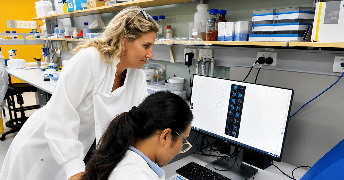
In Dr Samantha Barton’s laboratory, researchers have developed an advanced model using human stem cells to grow 'mini brains' in the lab. These mini brains contain myelin, and the team is a world leader in generating them.
This project aims to understand which genes are important for producing myelin in these mini brains. By identifying these genes, the team hopes to find new drugs that could promote myelin repair, with the long-term goal of helping to repair damage in people living with MS.
Multiple sclerosis (MS) is the most common acquired neurological disease in young Australians, affecting over 37,700 people. There is as yet no cure, and diagnosis often occurs years after symptoms begin.
Two key factors are strongly linked to the development of MS: infection with Epstein-Barr virus (EBV), a very common virus that infects over 90% of adults globally, and certain inherited immune genes known as HLA class II genes. However, it is still unclear how these factors interact to contribute to MS.
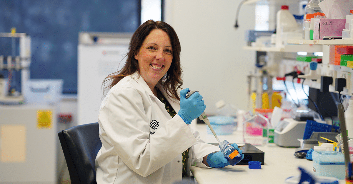
Dr Carla Proietti and her team have identified a specific pattern of EBV-related antibodies that is much higher in people living with MS than in people without MS or those with other autoimmune diseases. This pattern is also found in cerebrospinal fluid – the fluid surrounding the brain and spinal cord – suggesting that the immune response to EBV may be active in the brain in MS.
This project will investigate whether inherited HLA risk genes influence which EBV proteins the immune system targets, and whether this leads to harmful immune responses in the brain and spinal cord in MS. The team will analyse existing blood and cerebrospinal fluid samples from people with MS using advanced protein profiling technologies.
This research will help determine how genetic risk factors and EBV interact in MS. It may also identify specific viral targets linked to genetic risk, providing a clearer understanding of disease mechanisms. In the longer term, these findings could support the development of improved tools for MS diagnosis and disease monitoring.